Cataracts Case Study
General

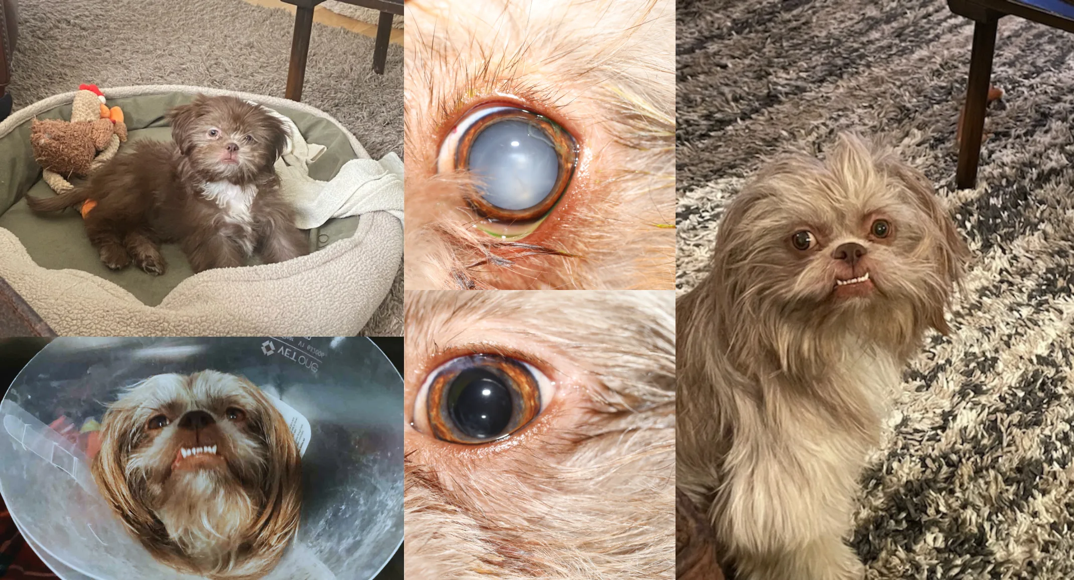
Charlie is a 2-year-old Shih Tzu that was recently diagnosed with bilateral cataracts. A cataract by definition is any opacity (cloudy in appearance) within the lens of the eye. Cataract surgery involves removing abnormal lens fibers using an ultrasound probe and replacing it with a prosthetic lens.
Charlie’s owners noticed that he was squinting his left eye in January of this year. They scheduled him to be seen by their regular veterinarian right away and they diagnosed him with bilateral cataracts. They noticed that the left cataract was significantly worse than the right, and that uveitis (inflammation inside the eye) was the cause of his squinting. They put Charlie on an anti-inflammatory eye medication (flurbiprofen) to reduce the inflammation and recommended that he come see us at AESH if they’d like more information about surgical removal of his cataracts.
Dr. Quantz, our ophthalmologist, saw Charlie at our hospital on 3/4/24 for a consultation to remove the immature cataract in his left eye. When a cataract is considered “immature”, it means that it involves more than 15-99% of the lens. A “mature” cataract involves the entire lens. Due to the degree of cataract formation in the left eye Charlie’s vision was compromised, but he maintained good vision in the right eye.
The ophthalmology team and the owners came up with a plan to surgically remove the cataract in Charlie’s left eye. Pre-surgical testing was the next step in moving forward and was scheduled for 2 weeks later. Shortly before this second visit Charlie’s owners noticed a significant decrease in his vision at home. By 3/18/24, it was noted that the cataract in the right eye had progressed significantly, leading to a reduction in vision. He was now running into walls and not able to find his ball while playing.
At Charlie’s recheck appointment, several tests were performed to ensure Charlie was a good candidate for surgery as well as general anesthesia. First, an ocular ultrasound was done to look for any anatomic abnormalities. Then, a gonioscopy was performed to assess whether he would be at an increased risk for increased pressure inside the eye after surgery. Finally, an electroretinogram showed that there were no abnormalities associated with the retina. In addition to these specific tests, we checked bloodwork, a urinalysis, tear production and ocular pressures.
Charlie returned on 4/9/24 for his cataract surgery. The procedure Dr. Quantz uses for cataract removal is called phacoemulsification. At AESH, we have a special machine just for this purpose. This machine generates ultrasonic waves through a small handpiece, to break up the cataract. This same handpiece will then aspirate (suck out) all the lens fragments while continuously maintaining globe inflation with irrigation fluids. This process was successfully completed by Dr. Quantz, and an artificial lens was implanted in each eye.
Like all surgeries, this procedure does have potential post-op complications. However, the success rate with this particular procedure is 80-90% within one year after surgery. This makes it the best option to fully restore vision for our cataract patients. Charlie’s owners will need to keep him quiet with an e-collar on for at least 4 weeks and he will need to stay on eye medications for the remainder of his life. The good news is that now he’ll be able to experience life with no visual barriers, just as he was meant to.
